Citizen / General public
Researcher
Student / Future student
Company
Public partner
Journalist
Teacher / Pupil

First in situ observations of metal biomineralization mechanisms at the bacterial cell level

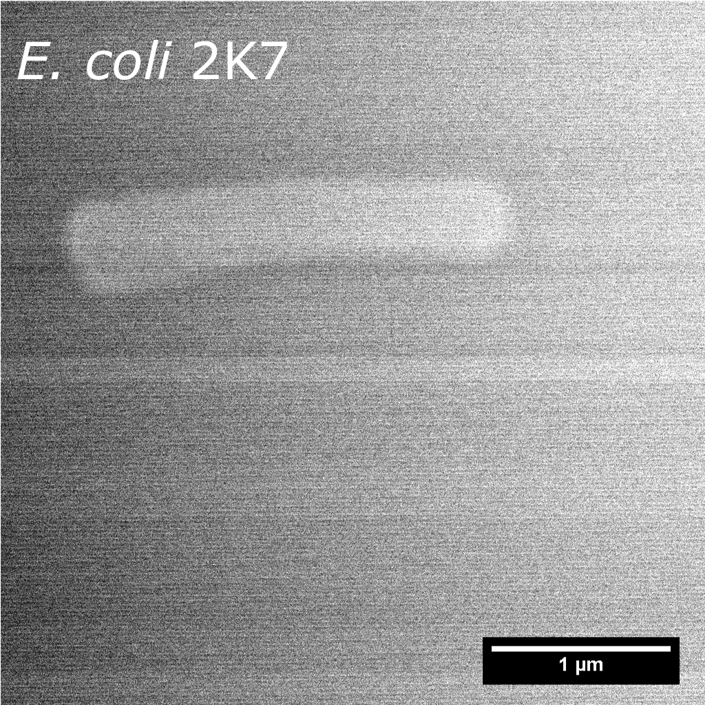
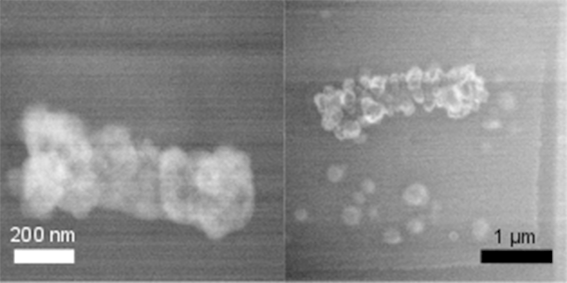
Scientists from the Institut de Physique du Globe de Paris and the Matériaux et Phénomènes Quantiques laboratory (University of Paris, CNRS), in collaboration with teams from the Muséum National d'Histoire Naturelle and the Institut Pasteur, have succeeded for the first time in characterizing, in a liquid medium and on a nanometric scale, the influence of bacterial cells and the organic polymers they secrete on the formation of a mineral.

First in situ observations of metal biomineralization mechanisms at the bacterial cell level

Publication date: 03/07/2020

Press, Research

Related themes : Origins

The nucleation and growth of the mineral depend closely on the nature and distribution of the functional groups within these structures, which ultimately determine the morphology, size and location of the mineral formed.

Biomineralisation is a process whereby minerals are synthesised by living organisms, in particular bacteria, which interact with metals in their environment, immobilising them in the form of solid minerals. This widespread phenomenon plays an important role in the cycling of metals in the environment, but until now it has never been possible to observe this process ‘live’.

In the environment, microorganisms are mainly organised in the form of biofilms, which are hydrated assemblies of microbial cells and organic polymers secreted by these cells. Among other things, this mode of organisation enables microorganisms to colonise the surface of minerals. These biofilms are also suspected of playing a major role in biomineralisation phenomena. However, the mechanisms governing the behaviour of metals within biofilms remain poorly understood to date because, to be representative of environmental processes, they need to be studied in hydrated conditions and at the scale of the microbial cell, i.e. submicrometre, which until now has been a major technological obstacle.

By taking advantage of recent advances in transmission electron microscopy in liquid cells, scientists from the Institut de Physique du Globe de Paris and the Matériaux et Phénomènes Quantiques laboratory (University of Paris, CNRS), in collaboration with teams from the Muséum national d’Histoire naturelle and the Institut Pasteur have succeeded for the first time in characterising, under hydrated conditions and on a nanometric scale, the influence of the nature and structure of bacterial cells and the organic polymers they secrete on the formation of a mineral.

Nucleation and growth of manganese precipitates on the bacterial cell envelope and in the extracellular medium. (IPGP/MPQ)

The team used two mutant strains of Escherichia coli, producing polymers of different natures, and induced manganese precipitation using the electron beam of the microscope. By then precisely locating the minerals formed, they showed that the initiation (or nucleation) and growth of manganese precipitates depend closely on the nature and distribution of the functional groups within these structures. The organic polymers thus act as supports that condition the morphology, size and location of the mineral formed.

Such in situ approaches on a nanometric scale open up new avenues for studying the major environmental processes that regulate the metal cycle in the critical zone. This constantly evolving surface envelope of our Earth is the site of complex interactions between rock, soil, water, air and living organisms that regulate the natural habitat and determine the availability of resources necessary for life, including our food production and the quality of our water.

Liquid transmission electron microscopy images of mutant E. coli strains secreting different types of organic polymers. On the left, the strain forming protein pili induces the formation of a manganese crust evenly distributed over its cell envelope. On the right, in the case of the strain producing cellulose on its surface but also in the extracellular medium, the manganese precipitates form more localised nanospheres. This variability in the distribution, size and morphology of the manganese minerals produced is linked to the nature and density of the charged functional groups, which vary according to the exopolymers produced by the bacteria (credits: IPGP/MPQ).

Ref : T. Couasnon, D. Alloyeau, B. Ménez, F. Guyot, J.-M. Ghigo, A. Gélabert, In situ monitoring of exopolymer-dependent Mn mineralization on bacterial surfaces. Sci. Adv. 6, eaaz3125 (2020).

Latest news
Jérôme Vergne becomes the new Director of the OVSM-IPGP
Committed to the various aspects of observing and monitoring telluric phenomena, Jérôme Vergne joined the IPGP on 1 July 2024 as Director of the Volca...
Evidence of magmatically induced faults at the East Pacific Rise
By comparison of ultra-high-resolution 3-D seismic imagery and bathymetry data collected at the East Pacific Rise (EPR) 9º50'N, researchers reveal the...
Charles Le Losq appointed member of the Institut Universitaire de France
The role of the Institut Universitaire de France (IUF) is to encourage the development of high-level research in universities and to strengthen interd...
NASA will measure earthquakes on the Moon using technologies developed for the InSight mission on Mars
The technology of the two seismometers that are part of NASA's Farside Seismic Suite instrument has detected more than a thousand earthquakes on the R...