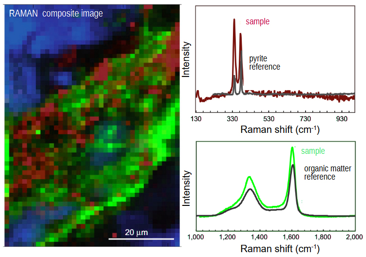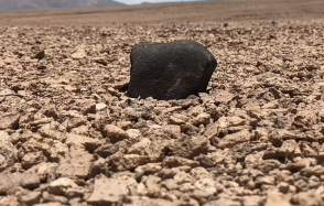
Discovery of CO₂ hydrates off the coast of Mayotte: a unique site for studying carbon storage in the ocean
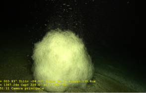
More than 120 CO₂ hydrate deposits were discovered at the Fer à Cheval site, 10 km east of Petite Terre (Mayotte), during the Geoflamme campaign co-le...