Afin d'être accessible au plus grand nombre, nous vous informons que vous avez la possibilité de modifier la taille d’affichage du contenu de notre site.
Pour cela, voici les commandes à reproduire à l'aide de votre clavier :
Zoom in
CTRL +
+
Zoom out
CTRL +
-
Reset to zero
CTRL +
0
Confocal laser scanning microscope coupled with confocal Raman microscope
Publication date: 20/03/2025
Research
These instruments can be used independently or in combination. The inViaTM confocal Raman microscope enables non-destructive imaging and characterization of any type of sample by Raman spectroscopy (surface imaging and analysis). The 2D or 3D imaging (enabled in confocal mode) is hyperspectral, i.e., all pixels in the images collected correspond to Raman spectra which can then be analyzed individually to characterize and locate compositional heterogeneities within the sample. The spectromicroscope is controlled and data are processed using WiRETM software (Renishaw). The instrument is equipped with a fast StreamlineTM (Renishaw) imaging system , which can collect up to 1,500 spectra per second to map large areas.
In practice:
● Spectral detection range from 130 to 4000 cm-1 ● Laser excitation at 514 and 785 nm ● Spectral resolution of 1 cm-1 ● Spatial resolution of 1 μm
The FV1000 confocal laser scanning microscope enables confocal observation of fluorescence via independent laser stimulation of biological or geological samples (with or without fluorescent probe labeling). Image stacks can be made in depth for 3D imaging, or in wavelength for spectral characterization.
In practice:
● 405, 488, 543 and 633 nm wavelength lasers ● High-speed imaging, 16 fps in 256 x 256 image format (4000 Hz) ● High-speed spectroscopy, 100 nm/msec ● Spectral resolution of 2 nm
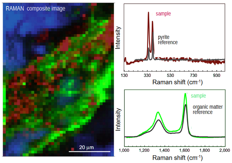
Caption: Thanks to the fast Raman imaging system (StreamlineTM system, Renishaw), installed on the inViaTM spectrometer, it is possible to generate 2D and 3D hyperspectral maps over large areas and to identify the various components and their distribution within the sample (here FeS2 pyrite and organic matter assemblages in 2.7 Ga stromatolites mapped using a 514 nm laser – in case of fluorescence in the sample, a 780 nm laser can be used; modified from Marin-Carbonne et al., 2018).
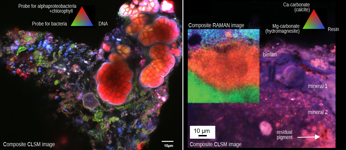
Caption: Confocal laser scanning microscopy (CLSM), implemented with an Olympus FV1000 microscope, allows the simultaneous imaging, using the 4 lasers available (405, 488, 543, and 633 nm), of naturally fluorescent components such as pigments or through labelling with specific or nonspecific probes (image on the left showing mineralized microbial mats; modified from Gérard et al., 2018). Minerals can be located by laser reflection or by combining Raman imaging (image on the right identifying and localizing minerals present within sections of modern microbialites embedded in resin; modified from Gérard et al., 2013).
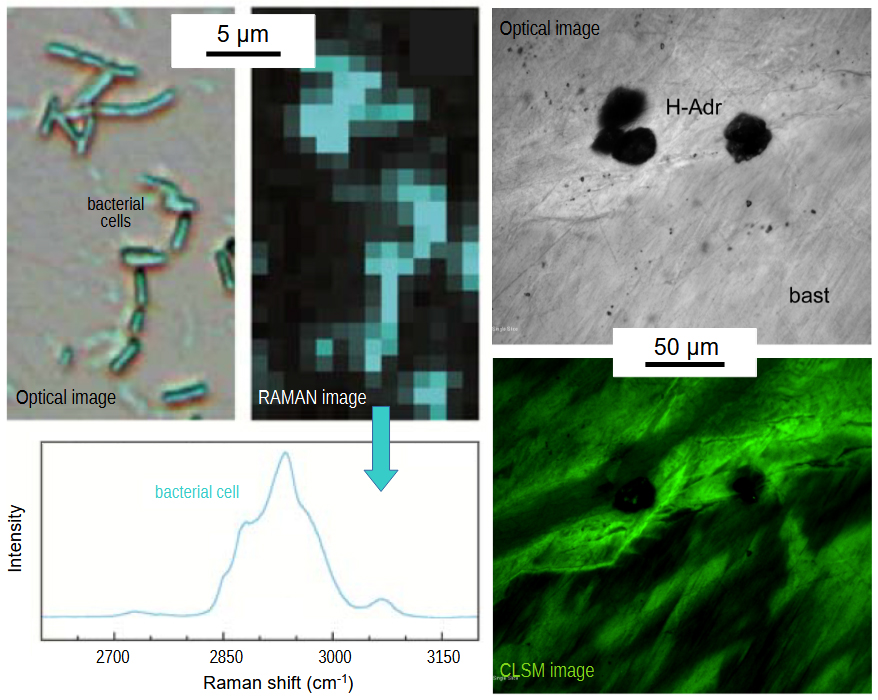
Caption: Raman and CLSM imaging can be implemented on a variety of objects (e.g., rocks, heritage or archaeological objects, materials, inorganic, organic and biological experimental products). Left: optical and Raman images of bacterial cells and associated spectra (modified from Ménez et al., 2012); right: optical and CLSM images of fluorescent organic compounds within rocks (modified from Pasini et al., 2013).
Planetary Research: a new open-access journal for planetary sciences
Led by an international community of researchers, with strong involvement from IPGP scientists, the new journal Planetary Research offers an alternati...
What if the next stars of recycling were… bacteria?
A new comic book from IPGP takes us behind the scenes of metal bio-recovery. Featuring bacteria, precious metals and a generous dose of humour, it rev...
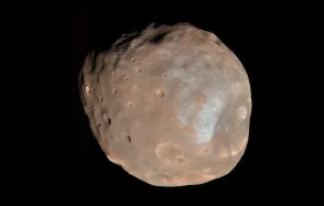
Mapping regolith migration on Phobos to support the MMX sample-return mission
Researchers from the Institut de Physique du Globe de Paris (IPGP), in collaboration with the Centro de Astrobiología (Spain) have published a new stu...
Lorraine Delaroque and Ines Ouar, two IPGP PhD students, receive awards at international conferences
Two doctoral students from the Planetology and Space Sciences team were recently recognized for their research at two international conferences.